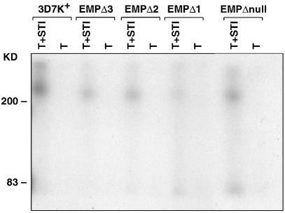
Fig. 7. Detection of PfEMP1 on the surface of infected erythrocytes by iodination and immunoprecipitation. Live 3D7k+, EMPΔ3, EMPΔ2, EMPΔ1 and the knockout parasite lines were surface labelled with 125I, SDS extracts made and labelled PfEMP1 immunoprecipitated with anti-ATS antisera. Before solubilization, the intact labelled cells were treated with either trypsin plus STI (T+STI) or trypsin alone (T) as described previously (Baruch et al., 1996).

An official website of the United States government
Here's how you know
Official websites use .gov
A
.gov website belongs to an official
government organization in the United States.
Secure .gov websites use HTTPS
A lock (
) or https:// means you've safely
connected to the .gov website. Share sensitive
information only on official, secure websites.
