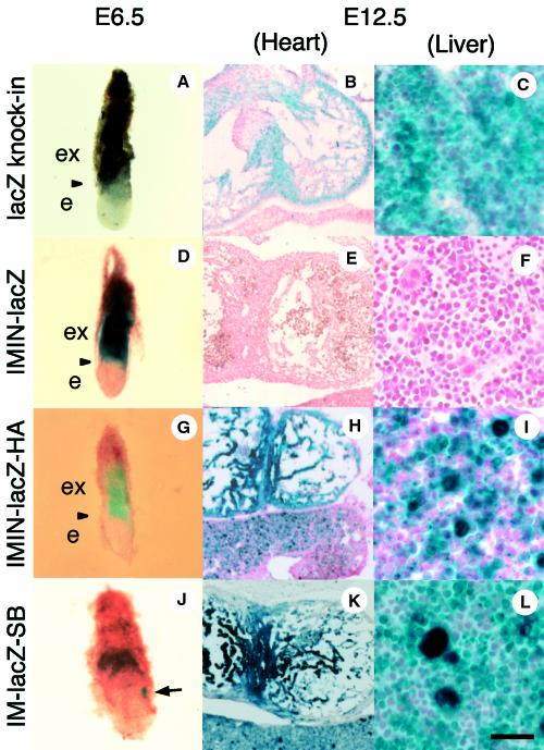
Fig. 3. Expression patterns of the lacZ knock-in and transgenic mouse lines. The ‘lacZ knock-in’ mouse (A, B and C) and IMIN-lacZ (line 880, D, E and F), IMIN-lacZ-HA (line 359, G, H and I) and IM-lacZ-SB (line 28, J, K and L) transgenic mouse embryos are shown. Embryos at E6.5 were processed for whole-mount staining (A, D, G and J), while older embryos were sectioned for detection of β-gal activity. Blue staining for β-gal activity was detected strongly in extra-embryonic tissue of the lacZ knock-in embryo (A) and IMIN-lacZ (D) and IMIN-lacZ-HA (G) transgenic embryos, whereas IM-lacZ-SB embryos showed blue staining only in the mesodermal region (indicated by an arrow in J). Heart and hematopoietic cells are stained strongly in E12.5 embryos of the lacZ knock-in (B and C), IMIN-lacZ-HA (H and I) and IM-lacZ-SB (K and L) transgenic embryos, but not in IMIN-lacZ embryos (E and F). Arrowheads in (A), (D) and (G) indicate the boundary between extra-embryonic tissue and the embryo proper. ex, extra-embryonic region; e, embryonic region. The scale bar corresponds to 180 µm (B, E, H and K) and 30 µm (C, F, I and L).

An official website of the United States government
Here's how you know
Official websites use .gov
A
.gov website belongs to an official
government organization in the United States.
Secure .gov websites use HTTPS
A lock (
) or https:// means you've safely
connected to the .gov website. Share sensitive
information only on official, secure websites.
