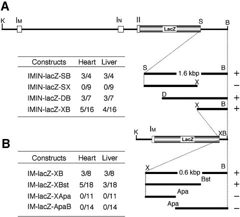
Fig. 4. Founder analysis of mafK 3′ regulatory sequences. All embryos were analyzed at E12.5, and liver and heart were examined for β-gal expression. (A) The structure of IMIN-lacZ-SB is shown on the top, which is a parent construct for IMIN-lacZ-SX, IMIN-lacZ-DB and IMIN-lacZ-XB. The truncated genomic fragments used to generate IMIN-lacZ-SB, IMIN-lacZ-SX, IMIN-lacZ-DB and IMIN-lacZ-XB are shown on the right. The incidence of β-gal-expressing embryos is shown to the left of the scheme of each construct. (B) The structure of IM-lacZ-XB is shown on the top, which is the parent construct for IM-lacZ-XBst, IM-lacZ-XApa and IM-lacZ-ApaB. The truncated genomic fragments used to generate IM-lacZ-XB, IM-lacZ-XBst, IM-lacZ-XApa and IM-lacZ-ApaB are shown on the right. The incidence of β-gal-expressing embryos is shown to the left of the scheme of each construct. The incidence is expressed as the number of β-gal-positive embryos/the number of transgene-positive embryos. The criteria for positive β-gal expression are blue staining in >20% of hematopoietic cells for the liver and either partial or complete staining of cardiac muscle cells for the heart. The constructs that retained enhancer activity are marked with +, and those without activity are marked with –. Apa, ApaI; B, BspHI; Bst, Bst1107I; D, DraI; K, KpnI; S, StuI; X, XbaI.

An official website of the United States government
Here's how you know
Official websites use .gov
A
.gov website belongs to an official
government organization in the United States.
Secure .gov websites use HTTPS
A lock (
) or https:// means you've safely
connected to the .gov website. Share sensitive
information only on official, secure websites.
