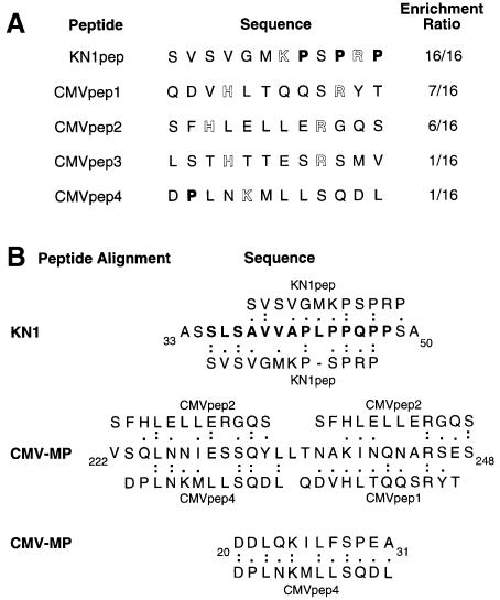
Fig. 1. Isolation and sequence analysis of plasmodesmal-interacting peptides. (A) A phage-display library was used in a screen to identify peptides that interacted with proteins in a plasmodesmal-enriched Nicotiana tabacum cell wall fraction (W2 fraction proteins). Proline residues have been indicated in bold type and basic residues are highlighted. (B) Amino acid sequence homology between identified phage-displayed 12mer peptides and motifs within the KN1 and CMV-MP. The bold type KN1 sequence represents the 14 amino acid oligopeptide, KN1-Npepsynth, used in microinjection experiments. (Single-letter code used for amino acid residues.)

An official website of the United States government
Here's how you know
Official websites use .gov
A
.gov website belongs to an official
government organization in the United States.
Secure .gov websites use HTTPS
A lock (
) or https:// means you've safely
connected to the .gov website. Share sensitive
information only on official, secure websites.
