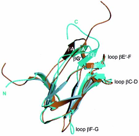
Fig. 6. The Runt domain complexed to CBFβ has a novel conformation. Least squares superposition of the Cα trace of the Runt domain crystal structure onto the most closely related NMR conformer [PDB code 1cmo, number 38, (Nagata et al., 1999)]. The analysis was performed for all the NMR conformers, and similar results were obtained. The NMR structure is shown in brown and the crystal structure is shown in cyan. The crystal and NMR structures differ in the relative positions of the βA′–βG′ sheet and the βF--G loop. The C-terminus is well defined in the crystal structure.

An official website of the United States government
Here's how you know
Official websites use .gov
A
.gov website belongs to an official
government organization in the United States.
Secure .gov websites use HTTPS
A lock (
) or https:// means you've safely
connected to the .gov website. Share sensitive
information only on official, secure websites.
