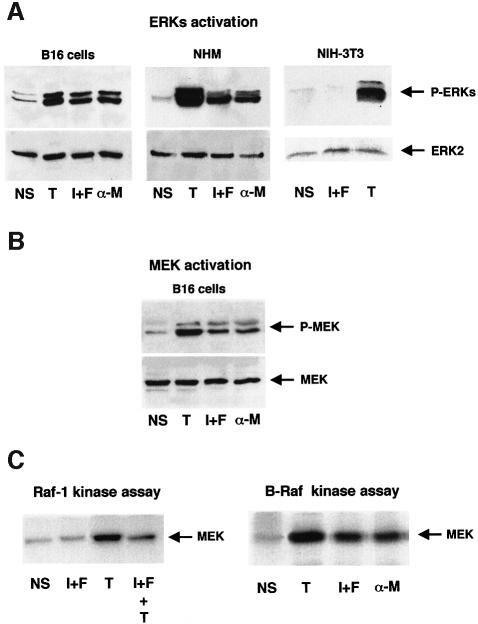
Fig. 1. cAMP activates ERKs, MEK and B-Raf in melanocyte. Lysates from non-stimulated cells (NS), cells stimulated for 10 min with the cAMP-elevating agents, IBMX plus forskolin (I+F) or α-MSH (α-M) or with the protein kinase C activator, TPA (T) were subjected to western blot analysis with an anti-phospho-ERK antibody (A) or with an anti-phospho-MEK1 antibody (B). A polyclonal antibody to ERK2 (1/2000) (A) or MEK (1/1000) (B) was used as a control of protein loading. (C) B16 cells were treated as above or pre-incubated for 20 min with cAMP-elevating agents and then stimulated for 10 min with TPA (I+F+T). B-Raf or Raf-1 was then immunoprecipitated and their kinase activities were assayed using the His–MEK fusion protein as substrate. Samples were resolved by 10% SDS–PAGE and analysed by autoradiography.

An official website of the United States government
Here's how you know
Official websites use .gov
A
.gov website belongs to an official
government organization in the United States.
Secure .gov websites use HTTPS
A lock (
) or https:// means you've safely
connected to the .gov website. Share sensitive
information only on official, secure websites.
