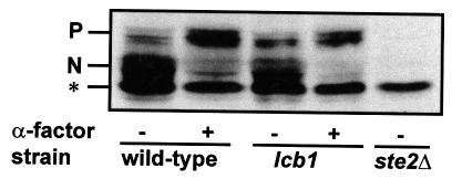
Fig. 6. Phosphorylation of the α-factor receptor. RH1800 (wild-type), RH3809 (lcb1) or RH3162 (ste2Δ) cells were incubated in the presence or absence of α-factor at 37°C. Total proteins were extracted and the α-factor receptor revealed by western blotting. N, normal phosphorylated α-factor receptor seen in the absence of pheromone; P, mobility of hyperphosphorylated form of the α-factor receptor after pheromone treatment; *, non-specific band seen in ste2Δ mutant.

An official website of the United States government
Here's how you know
Official websites use .gov
A
.gov website belongs to an official
government organization in the United States.
Secure .gov websites use HTTPS
A lock (
) or https:// means you've safely
connected to the .gov website. Share sensitive
information only on official, secure websites.
