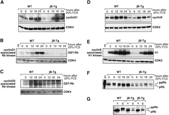
Fig. 5. Impaired cyclin D-associated kinase activity and reduced Rb phosphorylation in JunB-expressing 3T3 fibroblasts. Whole-cell extracts were prepared at the time points indicated post-serum stimulation from WT and jB-Tg 3T3 fibroblasts. (A) Western blot analysis of cyclin D1 and CDK4 expression levels. (B) Induction of cyclin D1-associated pRb kinase activity. The presence of CDK4 in the anti-cyclin D1 immunoprecipitate was confirmed by western blot analysis (ns, no substrate). (C) Induction of cyclin D2-associated pRb kinase activity. The presence of CDK4 in the anti-cyclin D2 immunoprecipitate was confirmed by western blot analysis. (D) Western blot analysis of cyclin E and CDK2 expression levels. (E) Induction of cyclin E–CDK2 kinase activity. The presence of CDK2 in the anti-cyclin E immunoprecipitate was confirmed by western blot analysis. (F) Western blot analysis of pRb expression and phosphorylation levels. A Molt-4 cell lysate was used as positive control (pc). (G) Western blot analysis of pRb expression levels in WT and jB-Tg 3T3 fibroblasts after serum starvation (s) or in an exponentially growing population (g). The results from two independent jB-Tg cell lines are shown.

An official website of the United States government
Here's how you know
Official websites use .gov
A
.gov website belongs to an official
government organization in the United States.
Secure .gov websites use HTTPS
A lock (
) or https:// means you've safely
connected to the .gov website. Share sensitive
information only on official, secure websites.
