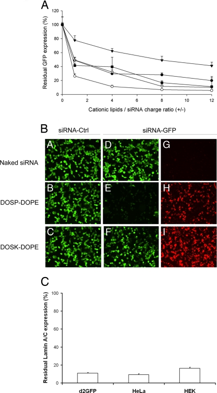
Fig. 4.
GFP silencing activity of the siRNA complexes formed by various aminoglycoside derivatives. (A) Residual GFP fluorescence after in vitro transfection of d2GFP cells with lipid/siRNA complexes characterized by various +/− charge ratios. GFP-expressing d2GFP cells were transfected with 400 ng of anti-GFP siRNA complexed with liposomes composed of BGTC (●), DOSK (▾), DOST (○), DOSP (■), and DOSN (▿). Residual GFP expression was expressed as the ratio (%) of GFP fluorescence in cells transfected with anti-GFP siRNA to GFP fluorescence in cells transfected with control (non-GFP targeting) siRNA. (B) Fluorescence microscopy visualization of GFP silencing and siRNA internalization. The GFP-expressing d2GFP cells were transfected with control siRNA (A–C in panel B) or 3′-rhodamine-labeled anti-GFP siRNA (D–I in panel B). The siRNA molecules (500 ng per well) were formulated in the absence (“naked” siRNA in A, D, and G in panel B) or in the presence of the cationic lipids DOSP (B, E, and H in panel B), or DOSK (C, F, and I in panel B). The transfected d2GFP cells were observed by using a FITC filter to visualize GFP fluorescence (A–F in panel B) or a rhodamine filter to visualize siRNA internalization (G–I in panel B). (C) Real-time quantitative RT-PCR analysis of human lamin A/C mRNA after transfection of various human cell lines (HEK293, HeLa, and d2GFP cells) with DOSP/siRNA lipoplexes [normalization to hypoxanthine–guanine phosphoribosyltransferase (HPRT1)]. Values are relative to cells transfected under the same experimental condition with a control siRNA.

