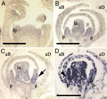
Fig. 3.
In situ expression pattern of IaTCP1 and IaH4 in young Iberis flowers. (A–C) IaTCP1 antisense probe hybridized to a longitudinal section through the Iberis inflorescence meristem and young developing flowers. (A) No distinct asymmetric IaTCP1 expression is detectable in the inflorescence meristem and adjacent young floral primordia (fp). (B) After onset of stamen differentiation, weak IaTCP1 expression becomes visible in petals (p) where similar expression levels were detected in adaxial (aD) and abaxial (aB) petals. Expression was also observed in anthers (lateral anther, la). (C) An asymmetric IaTCP1 expression becomes apparent in later floral stages, where a stronger signal was detectable in adaxial (arrow) than in abaxial petals. (D) Antisense IaH4 probe was hybridized to serial sections shown in C revealing that more cells express IaH4 in abaxial petals (arrowhead) compared with adaxial ones, which indicates higher cell proliferation activity in abaxial petals. (Scale bars: 200 μm.)

