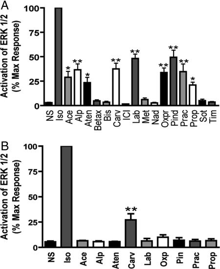
Fig. 2.
ERK activation in β2AR and β2ARTYY stable cells. HEK-293 cells stably expressing β2AR (A) or β2ARTYY (B) were stimulated with the panel of β2AR ligands used in Fig. 1 at 10 μM for 5 min, and cell lysates were analyzed for pERK and ERK by Western blot. pERK was normalized to total ERK protein. Data represent mean ± SE of at least three independent experiments done in duplicate. Quantification of pERK bands is as a percentage of maximal activity observed for isoproterenol. *, P < 0.05 vs. NS, **, P < 0.001 vs. NS.

