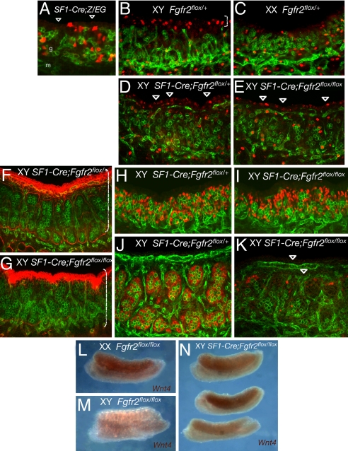
Fig. 3.
Abnormal testis development caused by conditional deletion of Fgfr2 by SF1-Cre in somatic progenitor cells. PECAM1 is green throughout. (A) Confocal scanning microscopy of a SF1-Cre/+;Z/EG gonad at 11.5 dpc shows a typical example of SF1-Cre-mediated activation of the Z/EG reporter (red) in a subset of gonadal somatic progenitor cells. Arrowheads indicate coelomic domain where few cells show recombination (compare with Fig. 2B). g, gonad; m, mesonephros. (B–E) Immunostaining and confocal scanning microscopy of a mitotic marker pHH3 (red) in controls and SF1-Cre;Fgfr2flox/flox gonads at 11.5 dpc. Somatic cells in coelomic domain show increased proliferation specific to XY gonads (bracket in B). (D and E) Proliferation in the coelomic domain (arrowheads) is only slightly decreased in XY SF1-Cre;Fgfr2flox/flox gonads relative to the normal levels in XY littermates. (F–K) There is little difference in gonad size between controls and mutant XY gonads at 12.5 dpc (brackets in F and G). (G) However, immunostaining of laminin (red) reveals aberrant testis cord formation in the mutant gonad. (I and K) SOX9 expression (red) appears normal in the mutant gonads at 11.5 dpc (I), but by 12.5 dpc it is severely reduced (K). PECAM reveals fragments of the testis vasculature (arrowheads in K). (L–N) Whole-mount in situ hybridization of Wnt4 in control and mutant gonads at 12.5 dpc. (L and M) In control gonads, Wnt4 expression is specific to XX gonads. (N) In XY SF1-Cre/+;Fgfr2flox/flox gonads, Wnt4 is activated but variable between samples and across the gonad field.

