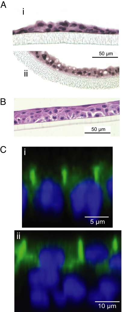
Fig. 2.
The morphology of mammary epithelial cells on Transwell permeable cell culture supports. (A) Sections through membranes of mPMEC in control medium (i) or lactogenic medium (ii), showing stimulation of lipid globules in lactogenic medium. (B) MCF10A cells grown on polyester membrane (untreated control). (C) Confocal images of mPMEC (i) and MCF10A (ii) cells immunostained for occludin (green) with nuclei (blue). Little or no occludin stain was observed in the basal layer.

