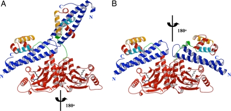
Fig. 2.
Overall structure of the TraRNGR–TraMNGR complex. (A) Structure of tetrameric NGR234 TraR–TraM complex. The TraRNGR–TraMNGR pair in the closed conformation is colored red and blue, respectively, whereas the other pair in the open conformation is in dark red and dark blue, respectively. The ligand AHL is shown in a ball-and-stick representation. α10, the major TraM-binding site, and α12, the DNA recognition helix, are colored in cyan and orange, respectively. The linker is colored in green. (B) Model of symmetric (TraRNGR–TraMNGR)2 in solution. The model was generated by applying the C2 rotational symmetry of the NTDs to the closed conformation of dimeric TraRNGR–TraMNGR. No structural conflicts are observed in the symmetric model. Views of A and B are the same. The figures were generated by using MOLSCRIPT and RASTER 3D (28, 29).

