
FIG. 3.
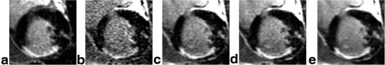
Short-axis images of the heart for a patient with inferior and inferolateral MI and a smaller anterolateral MI comparing (a) the “standard” breath-held, segmented IR-turbo-FLASH, (b) a single free-breathing, single shot IR-true-FISP image, (c) average of the first 8 repetitions free-breathing with motion correction, (d) average of the best 8 of 29 repetitions free-breathing with motion correction, and (e) average of all 29 repetitions free-breathing with motion correction.