
Figure 7. The dipole moments of the γ-crystallins in this study.
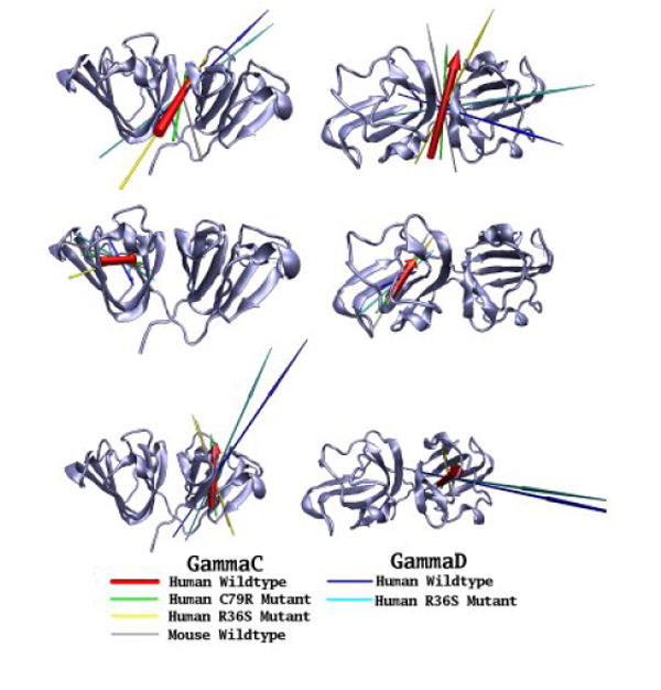
The dipole moments of six γ-crystallins (human wild-type γC, C79R, R36S and mouse γC; human wild-type γD and R36S mutant) have been calculated at the start of their simulation trajectories. Dipole moments of the whole protein (top line), N-terminal domain (middle line) and C-terminal domain (bottom line) are shown as arrows, with the length proportional to the magnitude, the underlying molecules have been superimposed using the group of residue concerned, with the secondary structure of the human wild-type γC-crystallins used as a reference point. Two views of the proteins are presented, from the ‘side’ and from the ‘top’ with the N-terminal domain on the left.