
Figure 8. The conserved ion pairs between motifs.
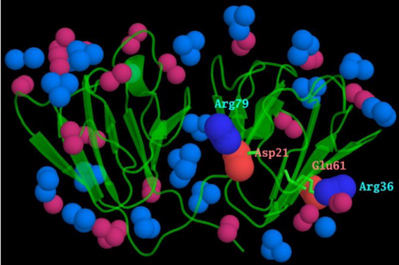
The surface of γ-crystallins is covered with ion pairs. Motifs 1, 2 and 4 have a conserved acidic and basic residue, highlighted in red and blue, in the alignment in Fig 1a. As a consequence of each domain being formed from motifs related by a pseudo-symmetrical twofold axis, these charged residues form inter-motif ion pairs. These are shown here, from the mouse γC-crystallin x-ray coordinates, with the N-terminal domain ion pairs between Asp 21 and Arg 79, and between Arg 36 and Glu 61 labelled. The atoms at the ends of the other acidic and basic side chains are shown in lighter colours. It can be seen that each of these inter motif ion pairs lies in the middle of a β-sheet, rather than at the “top” or “bottom” of the wedge-shaped domain where most of the other charged residues and ion pairs reside.