
Figure 4.
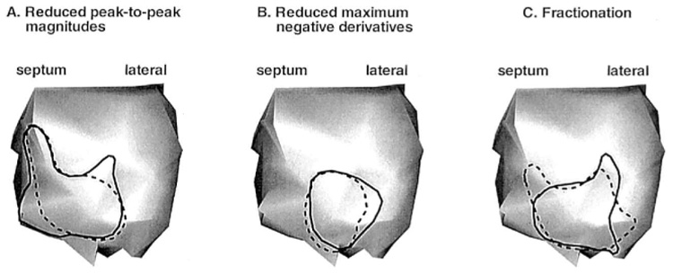
Estimated region of altered electrophysiologic substrate based on the posterior left ventricular (LV) endocardial pacing data of Figure 2 and Figure 3, using three different criteria. Solid lines depict estimates based on measured data. Dashed lines depict estimates based on reconstructed data. (A) Outlined LV regions contain electrograms with reduced peak-to-peak magnitudes (<15 mV). (B) Outlined LV regions contain electrograms with reduced maximum negative derivatives (<2 mV/msec, excluding superimposed sharp deflection). (C) Outlined LV regions contain fractionated electrograms.