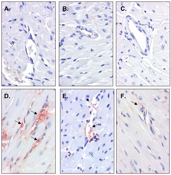
Figure 2.
Tumor-necrosis-α expression in myocardial tissue. Representative cross-sections of the myocardium showing immunohistochemical staining for TNF-α in mice administered: 0 LPS (placebo pellets) with either (A) 0 IF, (B) low IF (126 mg IF/kg diet), or (C) high IF (504 mg IF/kg diet); or LPS (pellets releasing 1.33 μg/d) with either (D) 0 IF, (E) low IF (126 mg IF/kg diet), or (F) high IF (504 mg IF/kg diet). Micrographs (20x) show no endothelial expression of TNF-α in the placebo mice but a marked increase in the animals receiving LPS (note arrows indicating TNF-α expression). TNF-α expression was down-regulated with increasing dose of IF.

