
Figure 5.
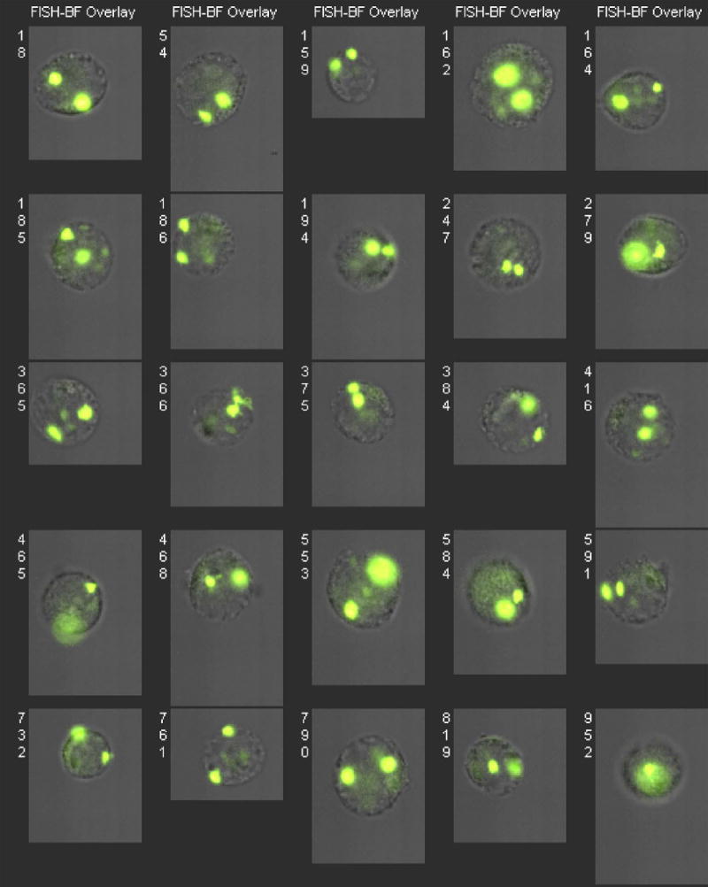
Jurkat Cells Hybridized in Suspension with a Chromosome 8 Probe and Imaged in Flow Using Standard Optics. Each cell is represented by a superposition of its chromosome 8 fluorescence (green) and brightfield (gray) images. Jurkat cells are larger than human PBMC and exacerbate variations in image focus quality.