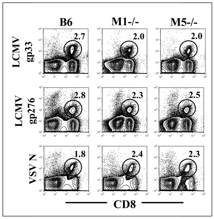
Figure 1. Visualization of anti-viral CD8+ T cells in mice with deletions of the M1 or M5 muscarinic receptors.

Mice were infected either with LCMV or VSV. Nine days later, splenocytes were stained with anti-CD8 antibody and tetramer specific for LCMV (gp33 or gp276) or VSV. Flow plots are gated on total lymphocytes and CD8+ tetramer + cells were visualized (shown in circles) The number within the plots reflects the percent of lymphocytes which bind the indicated tetramer.
