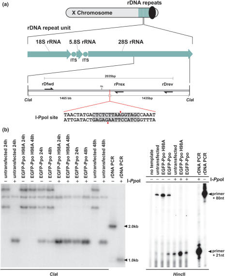
Figure 4.
I-PpoI expression and cleavage of genomic rDNA repeats in Sua 4.0 cell lines. (a) Schematic map of A. gambiae rDNA clusters and the location of the I-PpoI site within the 28S rDNA. ITS, internally transcribed spacer; Hc, HincII (HindII). (b) Southern blot analysis of A. gambiae Sua 4.0 cells transfected with pEGFP-Ppo or pEGFP-Ppo H98A. Genomic DNA was digested with ClaI and a 2 kb fragment of rDNA amplified with primers rDfwd and rDrev was used as probe. DNA in lanes marked by a (+) was digested with I-PpoI in vitro. (c) Primer extension analysis. Genomic DNA extracted from cells transfected with pEGFP-Ppo or pEGFP-Ppo H98A was digested with HincII and extended with primer rPrex. DNA in lanes marked by a (+) was digested with I-PpoI in vitro.

