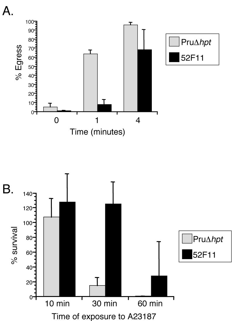
Fig. 1. IIE and IID phenotypes of PruΔhpt and the mutant 52F11 strain.
A. Intracellular parasites were exposed to 1μM A23187 Ca2+ ionophore for the indicated time period. Percentage egress represents the number of lysed vacuoles divided by the total number of vacuoles (lysed + intact). Each data point represents the average of three independent experiments and the error bars represent the standard deviation. At least 100 vacuoles were counted per experiment. B. Extracellular parasites were exposed to 1μM A23187 Ca2+ ionophore for the indicated time period before being added to cells. Percentage survival was calculated by dividing the number of plaques formed by parasites treated with A23187 divided by the number of plaques formed by untreated parasites. For each treatment all plaques in a well of a 24-well plate were counted. Data bars represent the mean of 3 independent experiments and the error bars represent the standard deviation.

