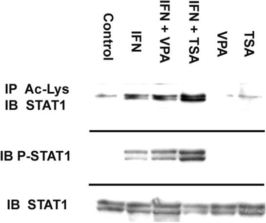
Figure 2. Immunoprecipitation of Ac-Stat1 from IFN-γ stimulated macrophages.

750 µg of extracted protein were mixed with 500 µl of PBS, 1 µl of normal mouse IgG and 20 µl of G-plus agarose. Anti-acetylated-lysine was added, followed by G-plus agarose. Protein was analyzed by western blotting with Stat1 p84/p91 antibody. In separate experiments, immunblots were performed for total Stat1 and phosphorylated Tyr701 Stat1 (P-Stat1). The cell lysates were separated by SDS-PAGE and transferred to PVDF membrane. Blocked membranes were then incubated with Stat1 p84/p91 or Tyr 701 Stat1 antibody. Blot is representative of three experiments.
