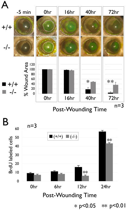
Figure 3. Wound healing is delayed in basonuclin-null corneal epithelium.
A, the upper panel shows representative images of the control (+/+) and basonuclin-null (−/−) eyes at different time points pre- and post-wounding. The images of –5 min show the eyes prior to wounding. The denuded surface of corneal regions was topically stained with fluorescein (green). In the 0 hr images, the denuded area is marked with a dashed line to illustrate the size of the original wound. The lower panel shows the quantification (n = 3) of the wounded area (presented as percentage of the original wound area). Note that the slower healing of basonuclin-null epithelium is observed at 40 hr and 72 hr post wounding, but not before. B, BrdU labeling showed a delayed onset of DNA replication in basonuclin-null corneal epithelium during wound healing. The bar graph represents the average number of BrdU labeled cells per section. Two sections from each of three mice were examined (n = 3). * p<0.05, ** p<0.01.

