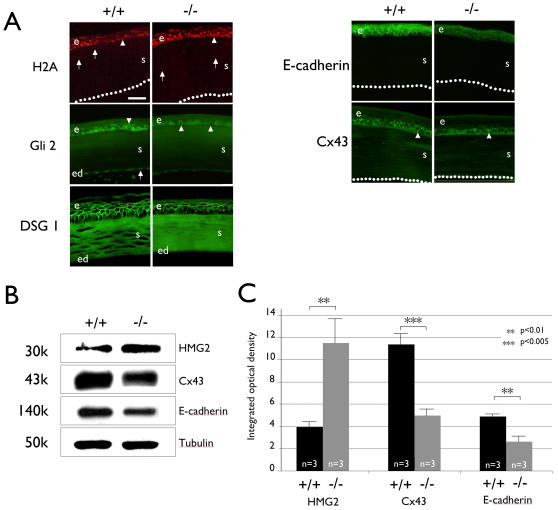
Figure 6. Basonuclin-null mutation affects level of proteins in basonuclin-targeted pathways.
A, Immunostaining shows perturbation of protein level in basonuclin-null corneal epithelium. The antibody specificity is indicated on the left of immunofluorescent images and the genotype on the top. Arrowheads show examples of a positively stained cell in the epithelium and arrows that in the stroma and endothelium. Dotted-lines indicate the surface of the endothelium. e, epithelium, s, stroma, ed, endothelium. B, Immunoblot of selected proteins whose mRNA were perturbed by basonuclin-null mutation. The antibody specificity is shown on the right of the blot image and the genotype on the top. The apparent molecular weight of each detected protein is given to the left. C, Quantification of the Immunoblot analyses. The values are average of three measurements (n = 3). **, p<0.01; ***, p<0.005.

