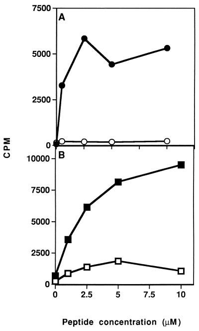
Figure 2.
Inhibition of in vivo priming of LN cells to the myasthenogenic peptides. (A) SJL mice were injected intradermally to the hindfoot pads with 10 μg of p195-212 in CFA, with ○ or without • concomitant i.p. inoculation of analog Lys-262–Ala-207 (200 μg in 500 μl of PBS). (B) BALB/c mice were injected intradermally to the hindfoot pads with 20 μg of p259-271 in CFA (100 μl), with □ or without ▪ concomitant i.v. inoculation of analog Lys-262–Ala-207 (200 μg in 500 μl PBS). LN cells taken from the mice 10 days later were incubated in the presence of various concentrations of the relevant peptide for 96 hr. Thereafter, [3H]thymidine was added and 16 hr later cells were harvested and radioactivity was counted. Results are expressed as mean cpm of triplicate cultures. SD values did not exceed 10%.

