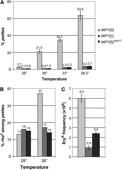
Figure 1.—
Mitochondrial DNA mutation frequency with different MIP1 alleles. DWM-5A-Δmip1, an haploid W303 derivative (Baruffini et al. 2006), was transformed with different MIP1 alleles. The MIP1 alleles were inserted into the SacI and SalI sites of the centromeric pFL39 plasmid (Bonneaud et al. 1991). The MIP1[S]A661T allele was produced by site-directed mutagenesis using the PCR overlap extension technique (Ho et al. 1989). (A) petite mutant frequency. Cells were pregrown at 28° on solid SC medium (6.7 g/liter yeast nitrogen base supplemented with a mixture of amino acids) supplemented with 2% ethanol. After 60 hr, the strains were replica plated on SC medium supplemented with 2% glucose and grown at the specified temperature. After 24 hr, strains were replica plated again on this medium. After 24 hr, cells were plated for single colonies on SC medium supplemented with 2% ethanol and 0.3% glucose. petite frequency was defined as the percentage of colonies showing the petite phenotype after 5 days at 28°. For each strain, at least 4000 clones were analyzed. Values are means of three independent experiments. (B) Percentage of rho0 mutants. The rho− clones containing mtDNA-deleted molecules and the rho0 clones devoid of mtDNA were distinguished as follows. At least 200 independent petite clones from each haploid mip1 strain were crossed with cox2, cox3, and two cob mit− mutants of opposite mating type on YPA plates (1% yeast extract, 2% bacto-peptone, and 40 mg/liter adenine) supplemented with 2% glucose, and after 2 days at 28° they were replica plated on YPA plates supplemented with 3% glycerol to identify rho+ diploids. In this work, a clone unable to complement any of the mit− mutants was arbitrarily defined as rho0. (C) EryR mutant frequency. Two independent series of 10 independent colonies grown on YPA plates supplemented with 3% glycerol were inoculated in 2.5 ml YPA medium. After 48 hr at 28°, 5–8 × 10⁷ cells were plated on YPAEG-ery medium (YPA supplemented with 3% ethanol, 3% glycerol, 3 g/liter erythromycin, and 25 mm potassium phospate buffer at pH 6.5) and grown at 28° for 9 days. An aliquot of each culture was plated for single colonies on YPA plates supplemented with 3% glycerol to determine the exact number of rho+ cells present in the culture.

