
Figure 1.
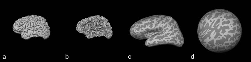
Cortical reconstruction. A given subject brain represented here by a 3D pial surface reconstruction (a) was transformed into gray matter/white matter interface surface (b), inflated (c) and transformed again into a spherical surface (d). Gyri and sulci are represented in light gray and dark gray colors, respectively.