
Figure 3.
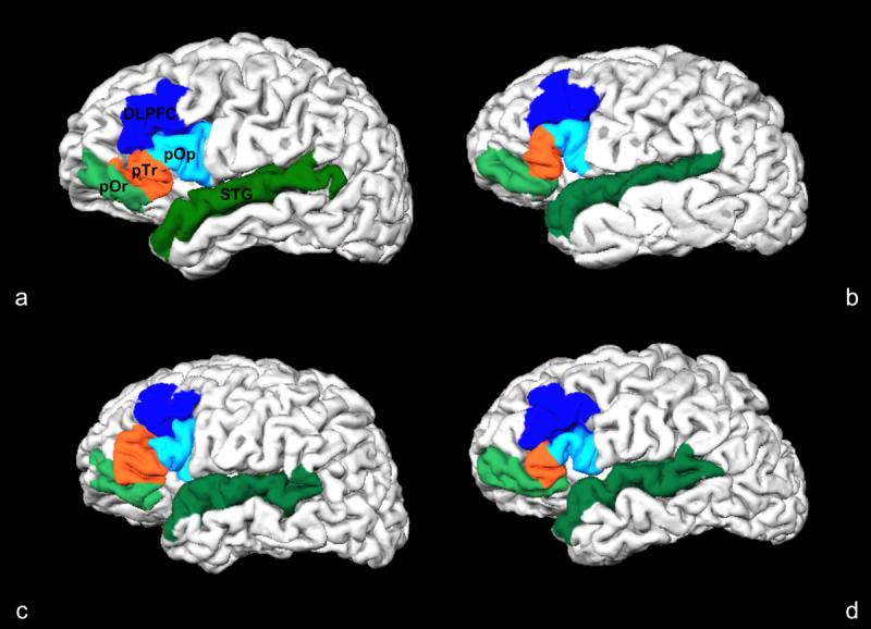
Cortical folding variability in Broca's area (pOr, pTr, pOp), dorsolateral prefrontal cortex (DLPFC) and superior temporal gyrus (STG). This illustrates the individual cortical folding variability of these ROI's between two control subjects (a, b) and two schizophrenia patients (c, d) in a pial surface reconstruction of brain image volumes. In the spherical registration of these subjects to an average subject template, the metric distortion required for optimal alignment is a reflection individual cortical displacement and convolution.