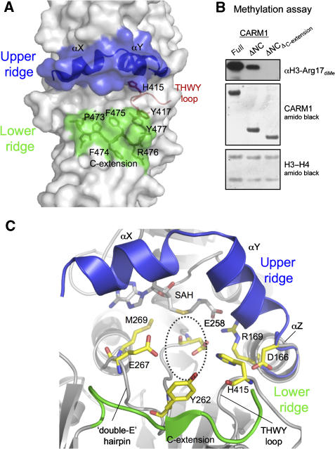
Figure 4.
CARM1 active site. (A) The active site forms a putative substrate-binding groove, lined by an upper ridge consisting of helices αX and αY (blue), and a lower ridge contributed by the C-extension PFFRY motif (green). The THWY loop forming part of the groove is also shown (red). (B) Methylation of histone H3-H4 tetramer by CARM1-full, CARM1-ΔNC or a ΔNC construct with the C-extension deleted ( ), probed by anti-H3 dimethyl-Arg17 antibody. (C) Conserved residues in the putative arginine pocket. The upper and lower ridges are coloured blue and green, respectively. This pocket can accommodate the side chain of the target arginine (position indicated by dotted circle).

