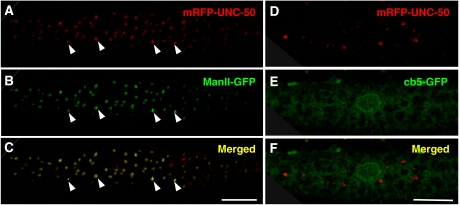
Figure 7.
UNC-50 localizes to the Golgi system. (A–C) mRFP-UNC-50 fusion proteins display a vesicular staining throughout the cytoplasm of a body-wall muscle cell, as shown. This largely overlaps the staining of the Golgi-resident Mannosidase II-GFP fusion protein. Representative Golgi structures are marked by arrowheads. (D–E) The mRFP-UNC-50 fusion protein does not overlap with an ER-localized GFP-cb5 protein. (The scale bar=10 μm.)

