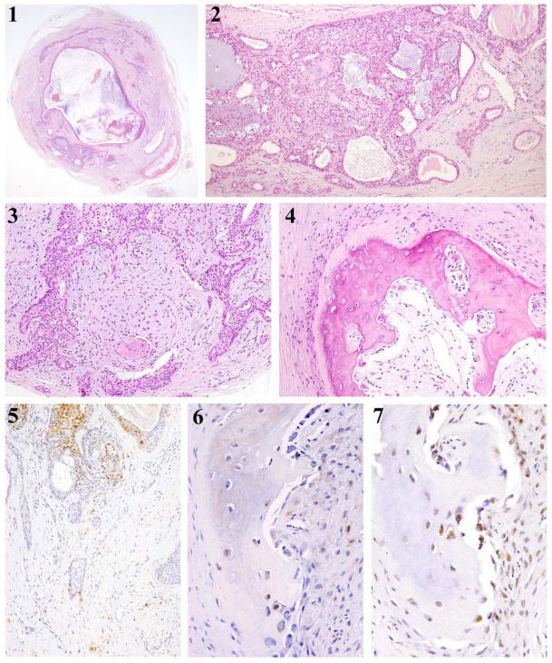
Figure 1.
(1) Tumor tissue is completely circumscribed with a fibrous capsule (HE, x 10). (2) The proliferating neoplastic cells consisted of ductal cells, sometimes showing formation of luminal formation with or without eosinophilic material (HE, x 30). (3) Spindle-shaped and oval-shaped neoplastic cells proliferating in so-called stromal tissues (HE, x 50). (4) Bone formation is observed in the tumor tissue. The bone has marrow-like tissue in the center of the bone tissue mass (HE, x 100). (5) S-100 positive products are detected in the neoplastic cells. Spindle-shaped or oval-shaped modified neoplastic myoepithelial cells react positively to S-100 protein (IHC, S-100, x50). (6) In some cases, S-100 positive products were detected in the cytoplasm of osteoblasts (IHC, S-100, x50). (7) The osteoblasts and osteocytes are both positive to Runx2 (IHC, Runx2, x50).

