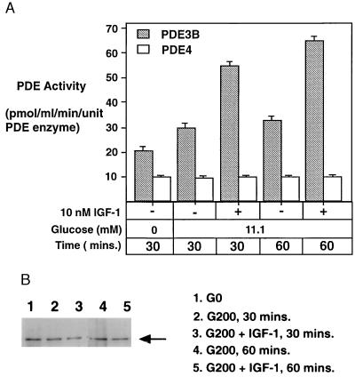
Figure 7.
Activation of PDE3B by IGF-1 in HIT-T15 cells. (A) When PDE3B activity was immunoprecipitated from the HIT-T15 extract, the pellet contained only PDE3B, as all of the PDE activity was completely suppressed by milrinone. A significant increase of PDE3B activity was observed when the HIT-T15 cells were treated with IGF-1. As a control, the PDE4 activity remaining in the supernatant did not change under any of the conditions tested. (B) Western blot assay indicating the integrity and relative amount of PDE3B protein present in each assay.

