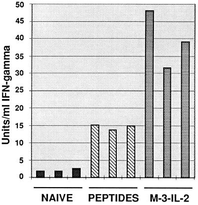
Figure 4.
IFN-γ release in response to M-3 target cells by nonadherent splenocytes from DBA/2 mice. Twenty-four weeks after metastases formation, splenocytes of disease-free mice cured by the IL-2-secreting cellular vaccine (M-3–IL-2) or fpLys–peptide mix treatment (Fig. 3a) were used in the assay. An age-matched naive mouse was used as negative control. Triplicate measurements of IFN-γ production are shown.

