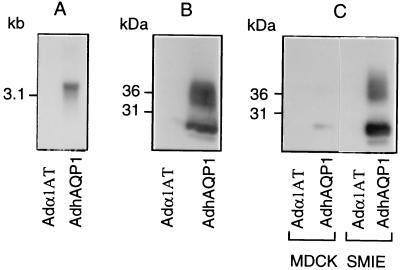
Figure 1.
hAQP1 expression in 293 cells and other epithelial cells. (A) Northern blot analysis was performed using 10 μg of total RNA, and 293 cells were infected at a MOI of 100 with either Adα1AT or AdhAQP1 and hybridized with a 813-bp radiolabeled probe of hAQP1 as described in Methods. (B) Ten micrograms of crude membranes from 293 cells infected at a MOI of 100 with either Adα1AT or AdhAQP1 were analyzed by Western blot as described in Methods. (C) Ten micrograms of crude membranes from MDCK and SMIE cells were analyzed by Western blot as above. The cells were infected at a MOI of 100 with either Adα1AT or AdhAQP1.

