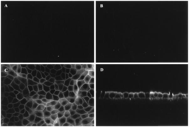
Figure 2.
Localization of hAQP1 in polarized MDCK cells. Confluent MDCK cells, grown on filters, were infected for 24 h at the basolateral side at a MOI of 100 with either Adα1AT (A and B) or AdhAQP1 (C and D). hAQP1 expression was determined as described in Methods. Micrographs of horizontal (xy; A and C), and vertical (xz; B and D) optical sections through MDCK cells are shown.

