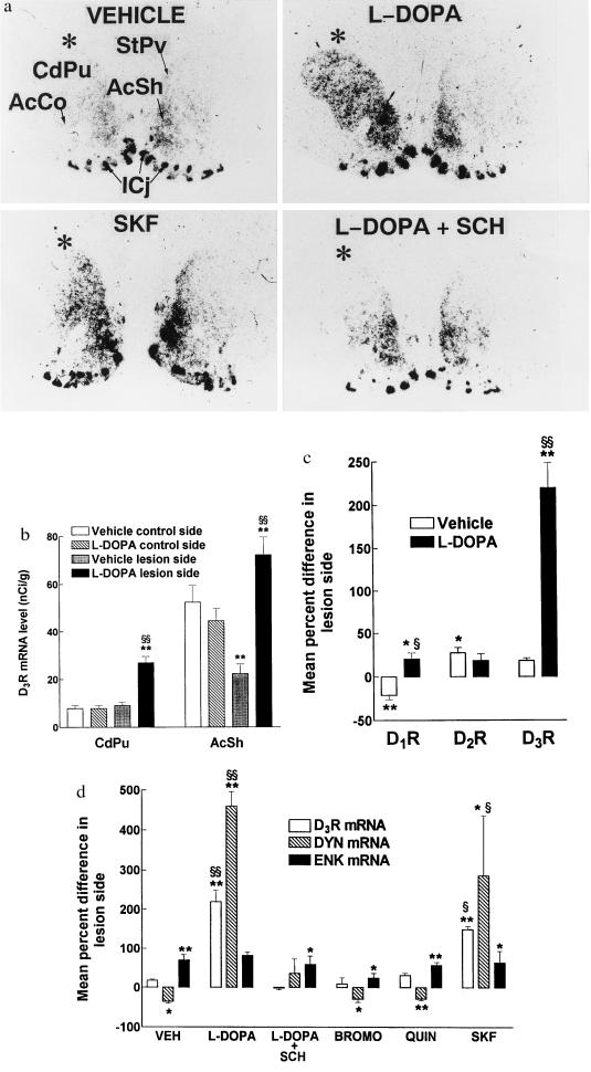
Figure 1.
In situ hybridization analysis of the effects of repeated administration of dopamine agonists on dopamine receptor and neuropeptide gene expression in the striatal complex of unilaterally 6-OHDA-lesioned rats. Coronal sections were hybridized with 33P-labeled cRNA probes for D1, D2, or D3 receptors (DRs), prodynorphin (DYN), or preproenkephalin (ENK) mRNAs, apposed to autoradiographic films and resulting pictures analyzed densitometrically. (a) Typical pictures showing the increased D3 receptor mRNA in the lesion side (indicated by asterisk) of the dorsolateral striatum (CdPu), StPv, and shell or core subdivisions of nucleus accumbens (AcSh, AcCo) after a 5-day levodopa (l-DOPA) treatment and the blockade of this effect by SCH 23390 (SCH). The effect of levodopa was partially reproduced by SKF 38393 (SKF). ICj, islands of Calleja. (b) Mean D3 receptor mRNA levels ± SEM (n = 4–6) in the two sides of CdPu (excluding StPv) and AcSh following repeated administration of vehicle or l-DOPA. (c) Mean percent difference (±SEM) in D1, D2, and D3 receptor mRNA levels between lesion and control side of CdPu. Same animals as in b. The mean (±SEM) signals on the control side of vehicle-treated rats corresponded to 454 ± 54, 597 ± 63, and 8 ± 1 nCi/g (1 Ci = 37 GBq) for D1R, D2R, and D3R, respectively. (d) Mean percent difference (± SEM, n = 4–6) in D3R, DYN, and ENK mRNA levels between lesion and control side of CdPu following 5-day administration of levodopa alone or together with SCH of bromocriptine (BROMO), quinelorane (QUIN), or SKF. The mean (± SEM) in situ hybridization signal on the control side of vehicle-treated rats corresponded to 0.22 ± 0.03 and 1.7 ± 0.2 μCi/g for DYN and ENK, respectively. ∗, P < 0.05; ∗∗, P < 0.01 in lesion vs. control side by the paired Student’s t test; §, P < 0.05; §§, P < 0.01 in lesion side of drug vs. vehicle-treated animals by the Mann–Whitney U test.

