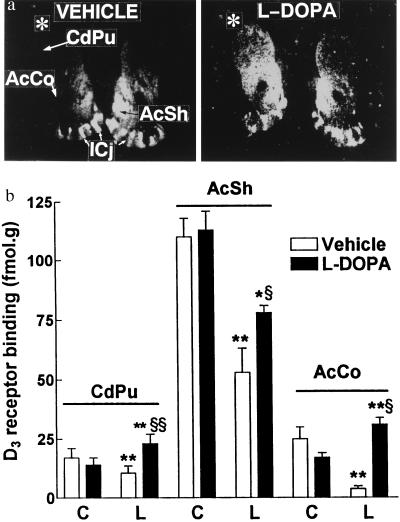
Figure 2.
Changes in D3 receptor binding elicited by repeated levodopa treatments of 6-OHDA-lesioned rats. (a) Digitized autoradiographic D3 receptor binding picture obtained with [3H]7-OH-DPAT, in animals treated twice a day for 5 days with vehicle (VEH) or levodopa (l-DOPA), in which nonspecific labeling has been subtracted. Asterisk indicates the lesion side. (b) Autoradiographic signals generated in a were quantified in CdPu, AcSh, and AcCo of control (C) and lesion (L) sides. Mean ± SEM of values from 4–6 animals. Nonspecific binding in CdPu was 50 and 15% of total in vehicle- and levodopa-treated animals, respectively. ∗, P < 0.05; ∗∗, P < 0.01 as compared with the control side by the paired Student’s t test; §, P < 0.05; §§, P < 0.01 in lesion side of drug vs. vehicle-treated animals by the Mann–Whitney U test.

