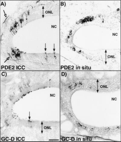
Figure 1.
PDE2 and GC-D are expressed in a subset of olfactory neurons. (A and C) Immunohistochemical localization of PDE2 and GC-D, respectively. (B and D) In situ hybridization localization of PDE2 and GC-D, respectively. The localization pattern of both proteins and mRNAs are similar. Open arrows denote PDE2 protein in axon fibers. Closed arrows point to cilia containing PDE2 or GC-D proteins extending from labeled neurons. NC, nasal cavity; ONL, olfactory neuron layer. (Bar = 50 μm.)

