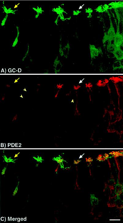
Figure 3.
Colocalization of PDE2 and GC-D in olfactory neurons. Confocal microscopy was used to determine that PDE2 and GC-D are found in the same neurons. (A and B) Unmerged image of olfactory neurons labeled with the GC-D and PDE2 antisera, respectively. (C) The merged image showing colocalization of the two proteins. PDE2 and GC-D are found in the same neurons. Some neurons, however, appear to express a lower level of PDE2 than GC-D (yellow arrow) in their cilia as compared with most PDE2/GC-D containing neurons (white arrow). Another unidentified cell type found throughout the neuroepithelium is also labeled by PDE2 antisera (arrowheads). (Bar = 10 μm.)

