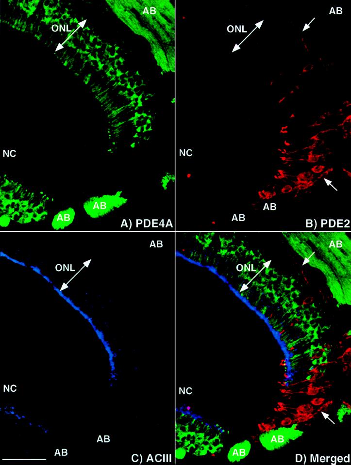
Figure 5.
PDE2 and GC-D are found in a subset of olfactory neurons distinct from those neurons expressing ACIII and PDE4A. Projection of a triple-labeled image demonstrating a unique subset of neurons containing PDE2: (A) PDE4A, (B) PDE2, (C) ACIII, and (D) merged image of A–C. None of these proteins is colocalized with another, indicated by no overlap of the signal upon merging of the images. ONL, olfactory neuron layer; AB, axon bundle; NC, nasal cavity. Arrow denotes axons expressing PDE2. (Bar = 50 μm.)

