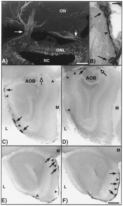
Figure 7.
Olfactory neurons that express PDE2 and GC-D project to a specific group of glomeruli in the olfactory bulb. (A) Projection of confocal images showing presence of PDE2 in axons forming bundles (arrows) and joining the olfactory nerve. (Bar = 50 μm.) (B) Two glomeruli (arrows) expressing PDE2 joined by labeled nerve fibers (arrowhead). (Bar = 200 μm.) (C–F) Serial sections (anterior-posterior) of the olfactory bulb demonstrating the pattern of glomeruli immunolabeled with PDE2 antisera (arrows). The modified glomerular complex is indicated by open arrows. Labeled nerve fibers are indicated by arrowheads. The labeled glomeruli “encircle” the caudal portion of the olfactory bulb. ONL, olfactory neuron layer; NC, nasal cavity; ON, olfactory nerve; M, medial; L, lateral. (Bar = 1 mm.)

