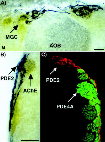
Figure 8.
PDE2 is found in glomerular complexes associated with high AChE activity (atypical glomeruli) but not to the same glomeruli that express PDE4A. (A and B) Olfactory bulb tissue sections were labeled with PDE2 antisera (blue) followed by an AChE activity assay (brown). The PDE2 signal is always associated with an area high in AChE activity. This is indicative of atypical glomeruli. (C) Olfactory bulb tissue was double labeled with PDE2 (red) and PDE4A (green) antisera. The signal appears in different glomeruli. MGC, modified glomerular complex; AOB, accessory olfactory bulb; M, medial. (Bar = 200 μm.)

