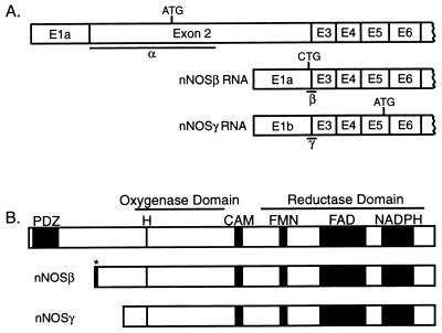
Figure 1.
Isoforms of neuronal NOS. (A) Illustration of mRNAs generated by alternative splicing of the nNOS gene. (Top) The predominant form in wt. Location of start codons and regions recognized by the nNOS α, β, and γ in situ probes are indicated. (Adapted from refs. 4 and 8.) (B) Illustration of nNOS isoforms. Consensus binding sites for heme (H), calmodulin (CAM), flavin mononucleotide (FMN), flavin adenine dinucleotide (FAD), and the reduced form of NADPH are indicated. nNOSβγ both lack the PDZ domain (PDZ), but the primary structure of the catalytic domains appears intact. ∗, six amino acids unique to nNOSβ. Based on refs. 14–16.

