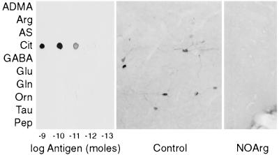
Figure 2.
Citrulline antiserum specificity. (Left) Dialyzed rat brain cytosol was coupled to various ligands with glutaraldehyde and then reduced with NaBH4. Serial 1:10 dilutions of each conjugate were spotted onto three pieces of nitrocellulose and then probed with a 1:10,000 dilution of antiserum. ADMA, asymmetric NG,NG-dimethylarginine; Arg, arginine; AS, argininosuccinate; Cit, citrulline; GABA, γ-aminobutyrate; Glu, glutamate; Gln, glutamine; Orn, ornithine; Tau, taurine; Pep, human glycine decarboxylase (residues 1,000–1,020). (Center) Mouse basal forebrain/islands of Calleja stained with 1:10,000 dilution of antiserum. Neuronal soma and processes are clearly labeled. (Right) Same area in mouse treated with nitroarginine, 50 mg/kg i.p. twice a day for 4 days. No neuronal staining is evident in this or other areas (data not shown) of the brain.

