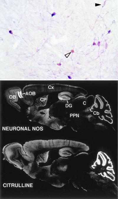
Figure 3.
Comparison of cit-IR and nNOS-IR. (Top) Double labeling for nNOS (purple) and citrulline (light brown-yellow). All cit-IR cells observed were also nNOS-IR, although many nNOS-IR cells devoid of cit-IR were observed (solid arrowhead). Cit-IR was especially high in the soma (open arrowhead), while nNOS-IR appeared both in the soma and processes. (Bottom) nNOS-IR and cit-IR in sagital sections of adult mouse. White areas represent positive staining. OB, olfactory bulb; AOB, accessory olfactory bulb; CP, caudate-putamen; DG, dentate gyrus; Cx, cerebral cortex; C, colliculi; PPN, pedunculopontine nuclei; Cb, cerebellum.

