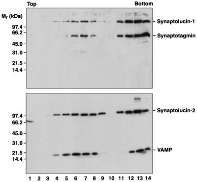
Figure 1.
Cofractionation of synaptolucins with synaptic vesicle proteins. Amplicon-infected PC12 cells were homogenized and postnuclear supernatants sedimented into 5–25% glycerol gradients. Small synaptic vesicles band in fractions 4–9, endosomes in fractions 11–14 (25). The bottom fraction contains material collected on a sucrose cushion; compared with the slower-sedimenting fractions, only 15% of this material was analyzed by SDS/PAGE. Proteins were precipitated with trichloroacetic acid, separated on 8–18% gels, and transferred to nitrocellulose. The filters were probed with mAb M48 (23), directed against synaptotagmin-I (Top), or mAb CL69.1 (24), directed against VAMP-2 (Bottom). Bound antibodies were visualized by ECL (Amersham).

