
Figure 4.
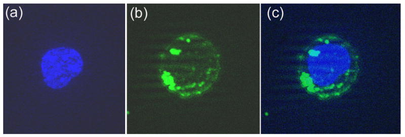
Fluorescence microscopy of the uptake of fluorescein-labeled PNAPBS-penetratin in CEM cells. The cells were incubated with fluorescein-labeled PNAPBS-penetratin conjugate for 30 min and then fixed and stained as described in the Materials and Methods. An aliquot of the cells was examined using the ZEISS Axiovert 200M microscope. Panel (a)-nucleus stained with DAPI; Panel (b)- cell cytoplasm showing bright green fluorescence of the fluorescent PNA; Panel (c)-nuclei merged with cell cytoplasm.