
Figure 2.
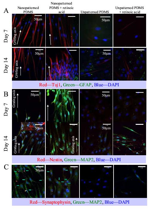
Immunofluorescent staining of (A) Tuj1 and GFAP, (B) MAP2 and nestin and (C) synaptophysin and MAP2 of hMSCs cultured on nano-patterned PDMS, nano-patterned PDMS in the presence of retinoic acid (RA), unpatterned PDMS and unpatterned PDMS in the presence of RA. (A) Tuj1 is shown in red, GFAP in green; (B) nestin is shown in red, MAP2 in green and (C) synaptophysin is shown in red and marked by arrows, while MAP2 is shown in green. In all panels the DAPI nuclei counter-stain is shown in blue, bar = 50 μm. The direction of the gratings on the nano-patterned PDMS is indicated with a white arrow. Images were taken at representative areas of the samples.